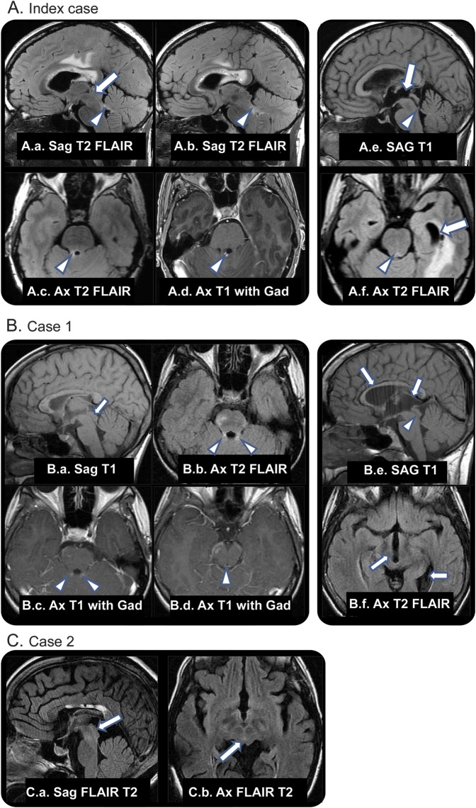
Figure. Cases.
(A.a–A.d) Neuromyelitis optica (NMO) lesions with normal aqueduct. (A.a) Normal caliber aqueduct (arrow). (A.a–A.d) T2 signal and gadolinium enhancement in NMO lesion of right superior cerebellar peduncle (arrowheads). (A.e, A.f) Eighteen months later, dilation of proximal aqueduct, third ventricle, and temporal horns (arrows) with stenosis or occlusion of distal aqueductal orifice (arrowheads). (B) Case 1. Chronic and enhancing NMO lesions, no ventricular obstruction. (B.a) Normal caliber aqueduct (arrow), chronic NMO lesions in corpus callosum. (B.b–B.d) T2 signal and enhancement in superior cerebellar peduncles and across the roof of the aqueduct (arrowheads). Additional enhancing focus in right cerebral peduncle. Thirty-two months later, symptomatic aqueductal stenosis. (B.e) Expansion of third ventricle and elevation of corpus callosum (arrows) and poor definition of aqueduct (arrowhead). (B.f) Dilation of third ventricle and expanded left ventricular atrium (arrows). (C) Case 2. MRI captured 11 years after initial shunt placement (earlier images were discarded). The absence of cerebral aqueduct within a small but otherwise normally developed mesencephalon supports the diagnosis of acquired aqueduct obliteration. (C.a, C.b) Absence of CSF flow void in expected location of aqueduct (arrows). Chronic changes in corpus callosum from NMO lesions. FLAIR = fluid-attenuated inversion recovery.

