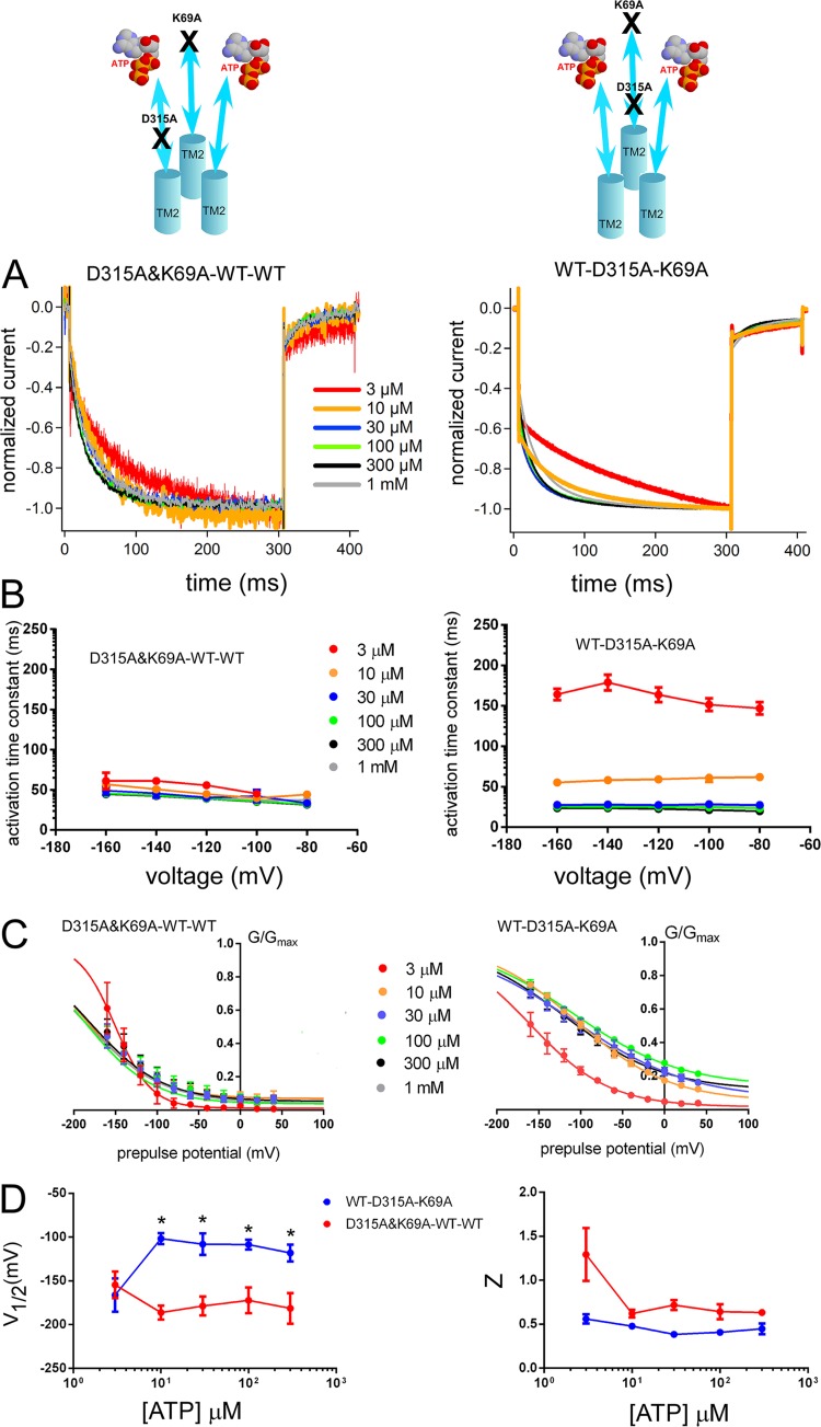
Figure 7.
The D315A linker mutation in one subunit shows different voltage- and [ATP]-dependent activation depending on the location relative to one ATP-binding site mutation (K69A) in the tandem trimer molecule. Analysis of D315A&K69A–WT–WT and WT–D315A–K69A shows different voltage- and [ATP]-dependent gating. (A) Normalized Voltage-induced activation traces at −160 mV for D315A&K69A–WT–WT (left) and WT–D315A–K69A (right) at various [ATP] from the representative data shown in Fig. S9. (B) Dependence of the activation time constants on voltage and [ATP] for D315A&K69A–WT–WT (left) and WT–D315A–K69A (right). Mean (± SEM) activation time constants for D315A&K69A–WT–WT (n = 6–10) and WT–D315A–K69A (n = 7–12) at various [ATP] and membrane potentials are shown. (C) Mean (± SEM) normalized G-V relationships at various [ATP] for tandem trimers D315A&K69A–WT–WT (n = 6–10) and WT–D315A–K69A (n = 6–16) derived from the maximum tail current responses at −60 mV by fitting with the two-state Boltzmann equation, as described in Materials and methods from the same oocytes. (D) Mean (± SEM) V1/2 (mV) and Z values for the D315A&K69A–WT–WT (n = 6–10) and WT–D315A–K69A (n = 6–16). *, P < 0.05.

