
Figure 3.
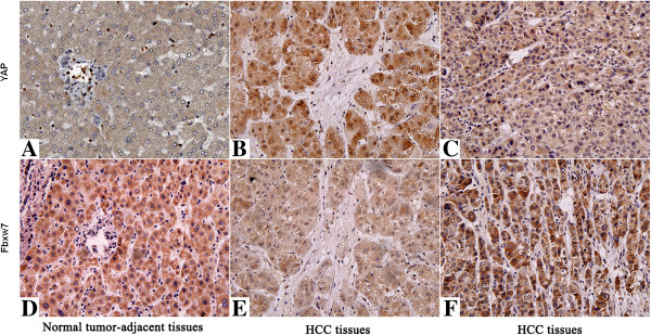
Immunohistochemical analyses of YAP and its correlation with Fbxw7 protein in HCC. In cases of high Fbxw7 protein expression (D, F), there was no detectable YAP protein expression (A, C) in the same tissue section. In contrast, in the case of low Fbxw7 protein expression (E), there was strong YAP protein expression (B). The fibrotic septa (B, E) simply represent fibrotic tumor stroma with normal background liver. Scale bar: 100 μm.