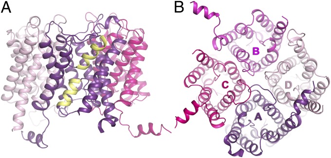
Fig. 1.
Overall structure of human AQP2. (A) Overview of the AQP2 tetramer with half helices formed by loops B and E highlighted in yellow. (B) Overview of the AQP2 tetramer from the intracellular side. The color scheme for each of the protomers is used throughout the article (A, purple; B, magenta; C, pink; D, light pink).

