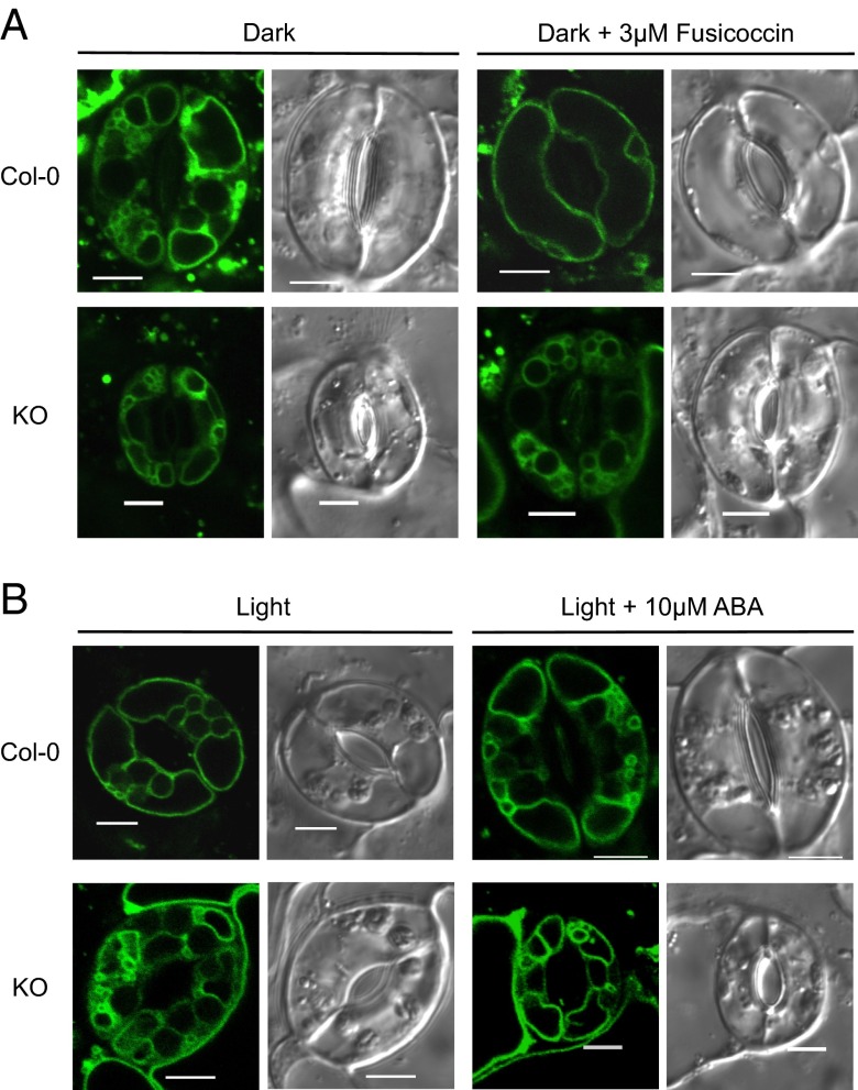
Fig. 5.
Vacuolar morphology of guard cells during stomatal movements. (A) Vacuolar structure of Col-0 and KO guard cells visualized with TIP1;1:GFP after dark incubation for 2 h (Left) and 3 µM fusicoccin treatment for 2 h (Right). (B) Vacuolar structure of Col-0 and KO guard cells visualized with TIP1;1:GFP after illumination for 2 h (Left) and followed by 10 µM ABA treatment (Right). Bright-field (Right) and GFP images (Left) of TIP1;1:GFP. (Scale bar: 5 µm.)

