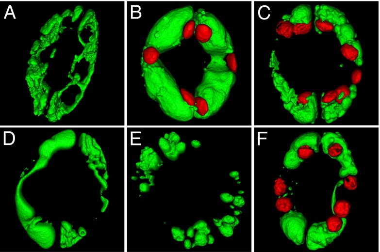
Fig. 6.
Three-dimensional projections of vacuolar morphology. (A) Surface rendering of guard cells vacuoles loaded with the BCECF-AM in closed stomata of WT plant. (B) Vacuolar morphology in open stomata of WT. Autofluorescence signal of chloroplasts was also captured and is shown in red. (C–F) Light-treated stomata of nhx1 nhx2 mutant plant. Chloroplasts are shown in red (C and F) or have been omitted (D and E).

