Fig. 4.
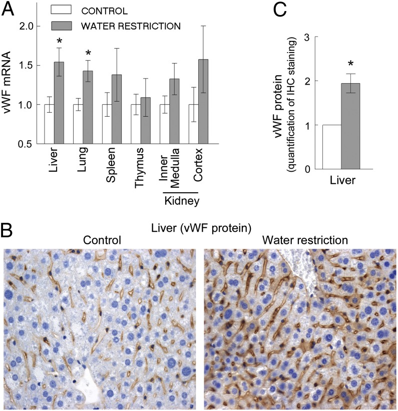
Water restriction increases vWF in endothelial cells of mice (mean ± SEM, n = 5, *P < 0.05, t test). Water was restricted for 9 d. (A) vWF mRNA increased in liver and lung. (B and C) Water restriction increased vWF protein in endothelial cells in the liver. (B) Representative images from immunohistochemical staining for vWF protein (brown) in the liver tissue sections. See Fig. S4 for pattern of blood capillaries in the liver (same sections stained for endothelial cells marker CD31). (C) Quantification of vWF in tissue sections. See Materials and Methods and Fig. S3 for details about image analysis.

