Fig. 2.

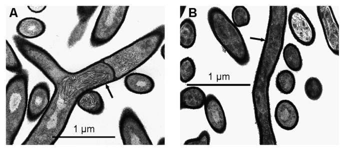
Electron microscopy images of Streptomyces scabiei EF-35 after 7 d of growth in minimal medium (A) or in suberin-supplemented medium (B), at a 35,590× magnification. Arrows show thicker cell wall in bacteria grown in the presence of suberin.

Electron microscopy images of Streptomyces scabiei EF-35 after 7 d of growth in minimal medium (A) or in suberin-supplemented medium (B), at a 35,590× magnification. Arrows show thicker cell wall in bacteria grown in the presence of suberin.