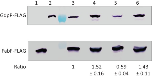
Fig. 4.

Detection of GdpP-FLAG and FabF-FLAG by immunoprecipitation followed by SDS-PAGE and Western blotting with anti-FLAG antibodies. The sections of the membrane containing GdpP-FLAG and FabF-FLAG were separated after antibody hybridization, and developed separately using a chromogenic assay. Strains used were: lane 1, fabF-FLAG (HB13056); 2, gdpP-FLAG (HB15845); 3, gdpP-FLAG fabF-FLAG (HB15857); 4, sigD gdpP-FLAG fabF-FLAG (HB15858); 5, flgM gdpP-FLAG fabF-FLAG (HB15859); 6, sigD flgM gdpP-FLAG fabF-FLAG (HB15860). The lane between lanes 2 and 3 is a protein ladder, where the 75 and 37 kDa markers are visible at the sections of GdpP-FLAG and FabF-FLAG proteins, respectively. This experiment was repeated three times, and one representative experiment is shown. The intensities of GdpP-FLAG bands were normalized with the internal protein control FabF-FLAG. The numbers below each band represent the fold change (mean±sem) of normalized GdpP-FLAG relative to strain HB15857 (lane 3).
