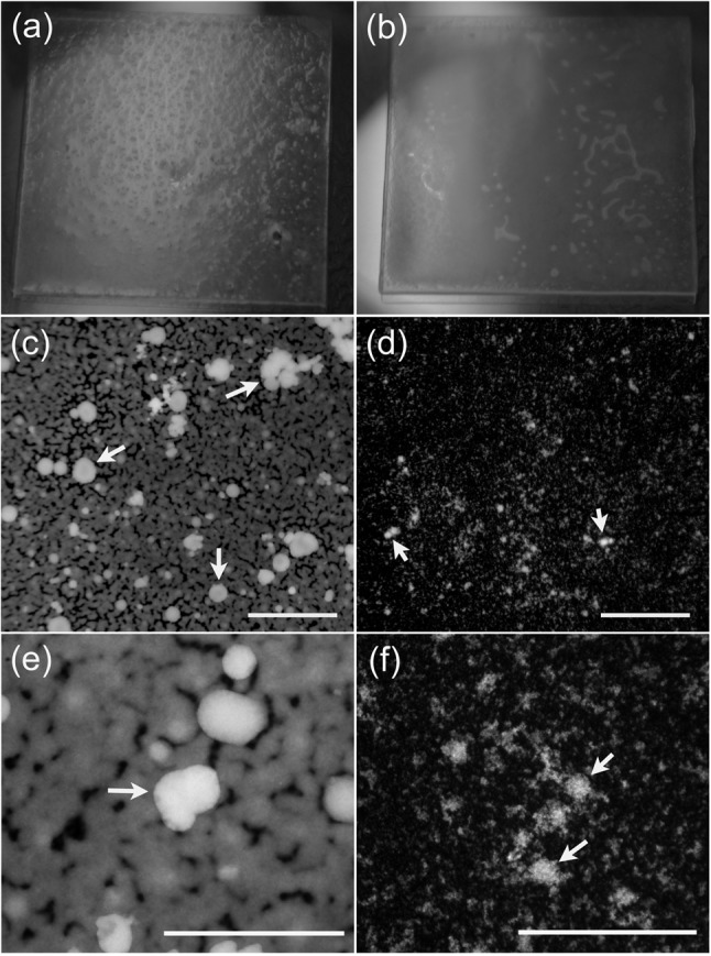
Fig. 2.

Structure of M. pneumoniae biofilms. (a, b) Macroscopic structure of a biofilm grown for 10 days on 18 mm ×18 mm glass coverslips prior to fixing. The biofilm formed by UAB PO1 (a) has a granular surface with numerous tower structures penetrating through to the surface of the biofilm, whereas that formed by M129 (b) has a smooth surface. (c–f) Fluorescence micrographs of the biofilms of UAB PO1 (c, e) and M129 (d, f) stained with propidium iodide. These low-power (×40) fluorescence images reveal the smooth margins of the biofilms in the honeycombed regions. The mycoplasmas between the towers are difficult to discern with the lower-power images of M129. (e, f) Higher-power (×400) images of biofilms formed by UAB PO1 and M129, respectively, revealing the towers and the honeycombed region of strain M129. Arrows indicate the towers. Bars, 400 µm (c, d) and 200 µm (e, f).
