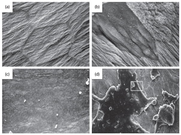
Fig. 2.
Urothelial alterations with bladder pathology. Scanning electron micrograph of apical surface of umbrella cell layer from (a) normal rat and (b) 2 h after spinal cord injury (reproduced from Apodaca et al.47 with permission). Scanning electron micrograph of hydro-distended bladders from (c) C-control (normal, unaffected) cats and in (d) cats diagnosed with feline interstitial cystitis (depicting regions where the umbrella cells layer is disrupted; reproduced from Lavelle et al.48 with permission).

