
Fig. 4.
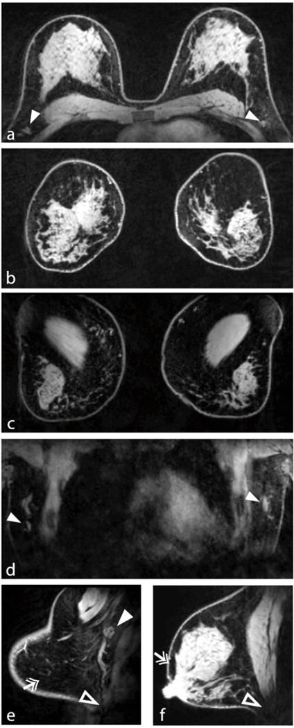
Demonstration of 7-T bilateral breast imaging. The transverse and reformatted coronal views (a, b) illustrate good coverage and uniform fat suppression. Coronal views through the chest wall (c) and anterior heart (d) demonstrate adequate penetration for visualisation of deep structures such as the pectoralis muscle and lymph nodes (solid arrowheads). Sagittal views demonstrate excellent fat suppression (double arrows) at the breast periphery (e) and near the nipple (f), which are areas that can be problematic owing to high susceptibility gradients. Signal at the intersection of the inferior breast and chest wall was limited because of a lack of coil coverage (open arrowheads in e and f)