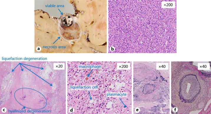
Fig. 3.
Macroscopic and pathological finding of the resected specimen. a The tumor consisted of viable and necrosis areas with well-demarcated nodular lesions at the caudate lobe (S1). The viable tumor size was 11 mm in diameter. b Histological examination showed a trabecular and pseudo-glandular structure with enlarged nuclei and hyperchromatins, which indicated moderately differentiated HCC in the viable area. c, d The necrosis area consisted of sclerotic fibrous stroma and liquefaction (arrows), and hyalinized degeneration (arrow) with hemosiderin-laden macrophages, plasmacytes and fibroblasts was found. e, f Vessel occlusion with organization (e), stenotic arteries with wall thickness (f) and mild chronic inflammation in fibrously enlarged portal areas were found in the necrotic area.

