
FIGURE 9.
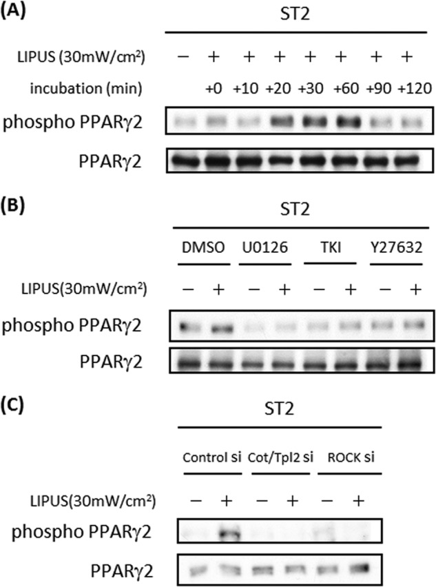
LIPUS induces PPARγ2 phosphorylation in ST2 cells. A, ST2 cells were cultured in adipogenic differentiation medium for 5 days. Cells were stimulated by LIPUS for 20 min and lysed in RIPA lysis buffer at the indicated time after the stimulation. Cell lysates were separated by SDS-PAGE, and levels of phosphorylated and total PPARγ2 proteins were determined by Western blotting. B, ST2 cells were pretreated with 5 μm Y-27632, 5 μm TKI, or 2.5 μm U0126 for 1 h followed by stimulation with LIPUS for 20 min. Western blotting analyses were performed as in A. C, ST2 cells were transiently transfected with Cot/Tpl2 siRNA, ROCK1 siRNA, or control siRNA (si). The analysis of PPARγ2 phosphorylation was performed by Western blotting as in B. mW, milliwatts.