FIGURE 2.
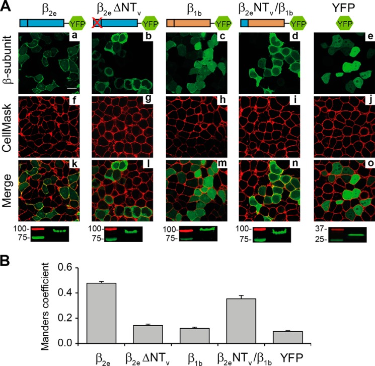
The Variable N-terminal segment of the β2e-subunit is responsible for targeting the protein to the plasma membrane. A, confocal fluorescence images of tsA201 cells expressing the indicated protein constructs fused to YFP as follows: full-length β2e (β2e); β2e lacking NTV (β2e ΔNTV); full-length β1b (β1b); chimeric protein encompassing NTV of β2e in a β1b background (β2e NTV/β1b). YFP moiety alone was used as control protein. Prior to visualization, cells were stained with the red plasma membrane marker CellMaskTM. Images from β2e fusion proteins, green channel (panels a–e); plasma membranes, red channel (panels f–j); and merge (panels k–o) are shown. Scale bar, 15 μm and is valid for all images. The bottom panels show crude lysates from cells expressing the indicated construct resolved by SDS-PAGE and visualized by fluorescence scanning. Numbers denote the molecular mass of standard proteins. The migration of the fusion constructs is consistent with their predicted molecular mass. B, bar plot of the colocalization analysis between β2e derivatives and the plasma membrane marker according to Manders coefficient. Values are expressed as mean ± S.E.

