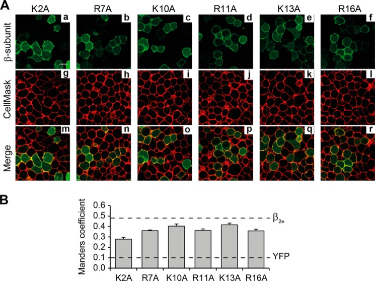
FIGURE 6.
Individual substitutions of positively charged residues within β2eNTv differentially alter plasma membrane targeting. A, confocal fluorescence images of tsA201 cells expressing the indicated β2e mutations at single lysine or arginine residues within β2eNTv. β2e single-point mutants (panels a–f), plasma membranes (panels g–l), and merged images (panels m–r) are visualized as in Fig. 2A. Scale bar represents 15 μm and is valid for all images. B, colocalization analysis and dashed lines as in Fig. 5B.

