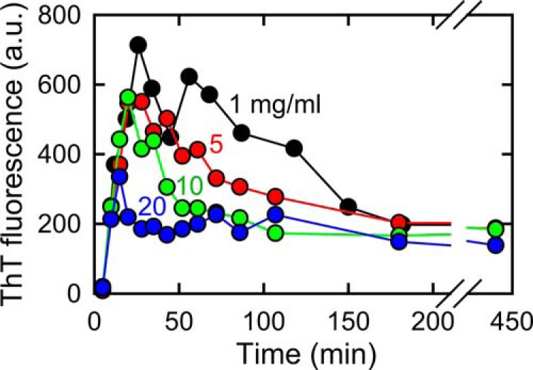
FIGURE 5.

Effect of protein concentrations on accumulation of the prefibrillar intermediate of insulin amyloid fibrils. Time course for the spontaneous formation of insulin amyloid fibrils in the presence of 1.0 m NaCl was monitored at 75 °C using ThT fluorescence at different concentrations of protein, i.e. 1 (black), 5 (red), 10 (green), and 20 mg/ml (blue). ThT fluorescence intensity was divided by protein concentration for plots of measurements at 5, 10, and 20 mg/ml of insulin to bring their level in line with that at 1 mg/ml.
