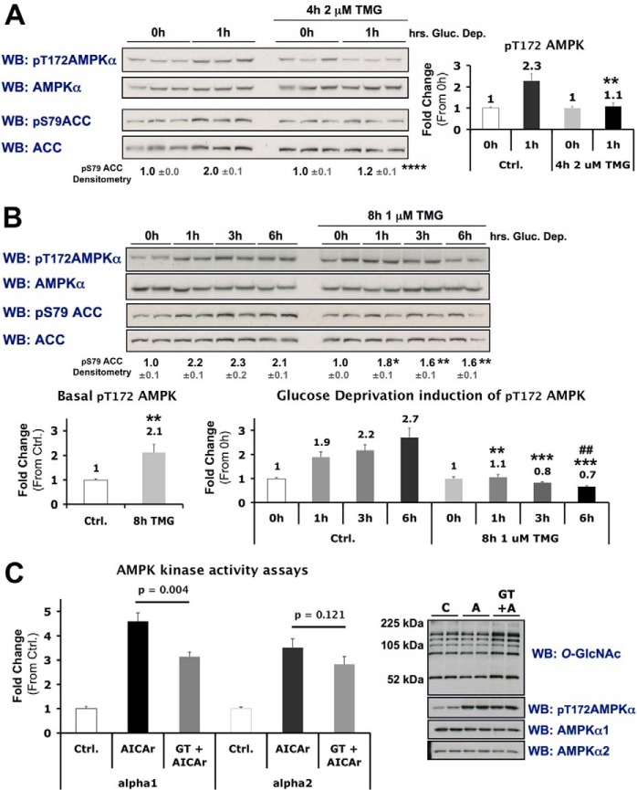
FIGURE 11.
Inhibition of O-GlcNAc cycling blunts activation of AMPK in differentiated C2C12 skeletal muscle cells. A and B, lysates from differentiated C2C12 cells incubated in 0 mm glucose for 0 or 1 h ± 4 h treatment with TMG (A), or 0, 1, 3 or 6 h ± 8 h treatment with TMG (B), were immunoblotted (WB) as indicated. Densitometric quantification of phospho- over total AMPK or ACC immunoblots are represented as graphs or bold numbers normalized to Ctrl., respectively. C, kinase activity assays were performed on AMPK-α1 and -α2 immunoprecipitates of lysates from differentiated C2C12 cells treated with vehicle (C or Ctrl.), AICAr (A; 1 mm, 30 min), or pre-incubated in GT (10 μm, 2 h) prior to AICAr treatment (GT+A). Right panels, representative control immunoblots of lysate used for kinase activity assays. All quantification represent mean values ± S.E. *, **, ***, and **** denote statistical significance of p < 0.05, p < 0.01, p < 0.001, and p < 0.0001, respectively, for TMG versus Ctrl for each time point. ## denotes a statistical significance of p < 0.01 when comparing the 0h versus 6h time points in TMG-treated cells.

