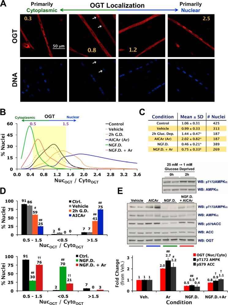
FIGURE 2.
Nuclear localization of OGT is tightly associated with AMPK activity in C2C12 myotubes. A, confocal projections of fixed C2C12 myotubes stained for OGT (red) and DNA (blue). Nuclear-to-cytoplasmic ratios of OGT immunofluorescence (NucOGT/CytoOGT) for each projection are indicated in yellow. A high degree of variability in nuclear localization of OGT within the same myotube is indicated with white arrows. B–D, quantification of NucOGT/CytoOGT values in C2C12 myotubes incubated for 2 h in fresh DMEM (Control or Ctrl.), serum-free DMEM (AICAr vehicle), 1 mm glucose DMEM (G.D.), AICAr (0.5 mm), a buffer deprived of all nutrients/growth factors except 25 mm glucose (NGF.D.), or 0.5 mm AICAr in NGF.D. buffer (NGF.D.+Ar). E, lysates from differentiated C2C12 cells exposed to the same conditions in parallel were immunoblotted (WB) as indicated. Mean NucOGT/CytoOGT and densitometric values of phospho- over total AMPK and ACC (±S.E.) are normalized to Ctrl. #, ##, and * denote statistical significance of p < 0.01, p < 0.0001, and p < 1 × 10⁻²⁸ versus Ctrl. and Veh., respectively. †, ††, and ‡ denote statistical significance of p < 0.01, p < 0.001, and p < 1 × 10−37 versus NGF.D., respectively.

