FIGURE 7.
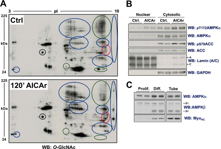
AICAr-induced activation of AMPK in differentiated C2C12 cells alters O-GlcNAcylated protein immunoblot patterning of the cytosolic fraction of lysates separated by 2DE. A, the cytosolic fraction of lysates from differentiated C2C12 cells treated with vehicle (Ctrl.) or AICAr (0.5 mm, 120 min) were separated by two-dimensional electrophoresis (2DE) and immunoblotted (WB) for O-GlcNAcylated protein. Blue, green, and red circles highlight regions where AICAr generally increased, decreased, or altered O-GlcNAcylated protein patterning, respectively. The black circle highlights a region that does not change, as reference for equal loading. B, control immunoblots, including lamin (A/C) (nuclear loading control) and GAPDH (cytosolic loading control), of the nuclear and cytosolic fractions of the same lysates presented in panel A confirms activation of AMPK. C, lysates from proliferating (Prolif.) and differentiated (Diff.) C2C12 cells, and lysate from myotubes enriched from differentiated C2C12 cells (Tube) were immunoblotted as indicated (myosin heavy chain (MyoHC; myotube marker)), confirming β2-subunit harboring AMPK complexes as the predominantly expressed isoenzyme in myotubes.

