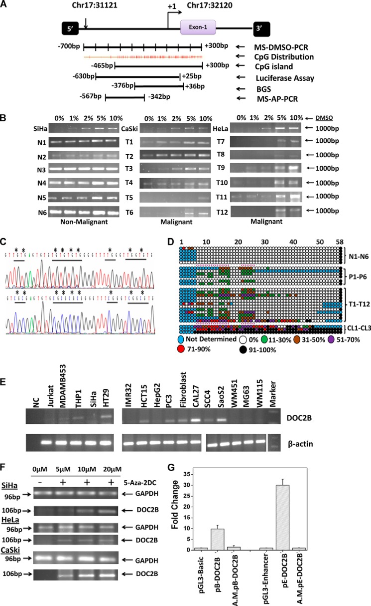
FIGURE 1.
Methylation profiling of the Frag-13 fragment in cervical samples. A, schematic representation of the region selected for validation of Frag-13 fragment by MS-AP-PCR, MS-DMSO-PCR, BGS, and characterization of the promoter region of the DOC2B gene by a luciferase assay. B, MS-DMSO-PCR showed that DNA methylation changed the sensitivity of amplification to the DMSO concentration in the PCR mixture. In the case of methylated samples, amplification was detected at a DMSO concentration of 2% or more, whereas unmethylated or less methylated samples showed amplification even in the absence of DMSO. N and T represent non-malignant and tumor samples, respectively. C, representative electropherogram of a portion of the 412-bp region of non-malignant and tumor samples showing methylated and unmethylated CpG sites. The differentially methylated CpG sites were highlighted by asterisks (*). D, methylation map of the DOC2B promoter fragment in non-malignant, pre-malignant, malignant, and cervical cancer cell lines. The open and filled circles represent the unmethylated and methylated CpG sites, respectively. The partially methylated CpG sites were filled with different colors depending on the extent of methylation. Each horizontal line represents single samples, and circles representing single CpG sites. N1-N6, P1-P6, T1-T12, and CL1-CL3 represent 6 non-malignant, 6 pre-malignant, 12 malignant cervical samples, and 3 cervical cancer cell lines namely SiHa, CaSki, and HeLa, respectively. E, expression analysis of DOC2B in various cell lines by semi-quantitative RT-PCR with β-actin as control. F, demethylation by 5-aza-2DC and subsequent reactivation of mRNA of the DOC2B gene. Cervical cancer cell lines SiHa, CaSki, and HeLa were used for demethylation and reactivation experiments. In all three cell lines, reactivation was observed at 5 μm or above of 5-aza-2-DC treatment. GAPDH was used as an internal control for the integrity of the cDNA. RT-PCR analysis shows the loss of DOC2B expression and treatment with the demethylating agent 5-aza-2-DC restores its expression. G, characterization of promoter activities of the DOC2B gene by transient transfection experiments using DOC2B promoter constructs in pGL3-Basic and pGL3-Enhancer vectors. Histograms represent mean ± S.D. for at least two independent experiments. pB-DOC2B and pE-DOC2B represent the DOC2B promoter constructs cloned in pGL3-Basic and pGL3-Enhancer vectors, whereas A.M.pE-DOC2B represents corresponding artificially methylated constructs as discussed under ”Experimental Procedures.“

