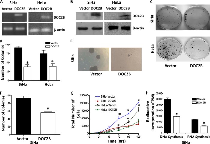
FIGURE 2.
Effect of ectopic expression of DOC2B on cell growth and proliferation. A and B, representative figures showing expression of the DOC2B gene upon transfection by RT-PCR and Western blot, respectively. DOC2B was detected by anti-DDK tag antibody. β-Actin was used as an internal control. C, DOC2B inhibits tumor growth in vitro in SiHa and HeLa cells, respectively. D, quantitative analysis of colony forming assay represented as mean ± S.D., *, p < 0.05, shows a significant decrease in colony number after ectopic expression of DOC2B. E, representative image of soft agar colony forming assay. F, quantitative analysis of colony number represented as mean ± S.D.; *, p < 0.05. G, represents the cell proliferation rate in DOC2B expressing stable clones in comparison with vector control. Ectopic expression of DOC2B significantly inhibited cell proliferation resulting in delayed cell doubling time. The cell doubling analysis was performed using the cell doubling time calculator. H, cell proliferation rate was significantly inhibited at both DNA and RNA levels in DOC2B expressing cells when compared with control cells by [3H]thymidine and [3H]uridine incorporation assays, respectively (mean ± S.D. from 3 independent experiments in duplicates). *, represents p < 0.05.

