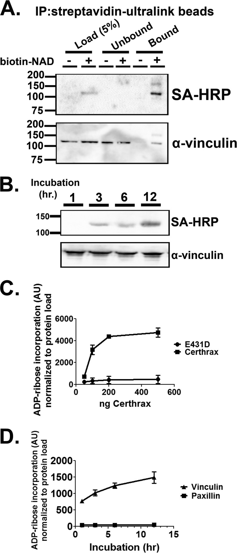
FIGURE 1.
Precipitation of biotin-NAD-labeled substrate with immobilized streptavidin pulls down vinculin protein. A, HeLa lysate was incubated with Certhrax and biotin-NAD for 1 h. Biotinylated protein (ADP-ribosylated) was then immunoprecipitated with Streptavidin-Ultralink beads. The beads were washed with TBS and bound material was eluted with free biotin and SDS. The reactions were resolved by SDS-PAGE and transferred to PVDF membrane. Immunoblot with α-vinculin or streptavidin-HRP (SA-HRP) conjugate detected vinculin and biotin-ADP-ribose, respectively. B, recombinant vinculin purified from E. coli was incubated with CerADPr and biotin-NAD for the indicated times. The reaction was stopped by addition of Laemmli buffer and boiling. The reaction was resolved by SDS-PAGE and transferred to PVDF. Biotin-ADP-ribose was detected with streptavidin-HRP conjugate. Total vinculin was detected by α-vinculin immunoblot. C, recombinant vinculin was incubated with increasing doses of CerADPr or catalytically inactive CerADPr(E431D). After 3 h, the reaction was stopped by addition of Laemmli buffer and boiling. The reaction was resolved by SDS-PAGE and transferred to PVDF. Biotin-ADP-ribose was detected with streptavidin-HRP conjugate and total protein was detected with α-vinculin antibody. Biotin signal was normalized to total vinculin (VCL) signal and expressed in arbitrary units (AU). D, recombinant vinculin or recombinant paxillin were incubated with CerADPr for the indicated times. At each time point, an aliquot was removed and the reaction was stopped by addition of Laemmli buffer and boiling. The reaction was resolved by SDS-PAGE and transferred to PVDF. Biotin-ADP-ribose was detected with streptavidin-HRP conjugate and total protein was detected with α-vinculin antibody. Biotin signal was normalized to total protein load and expressed in arbitrary units.

