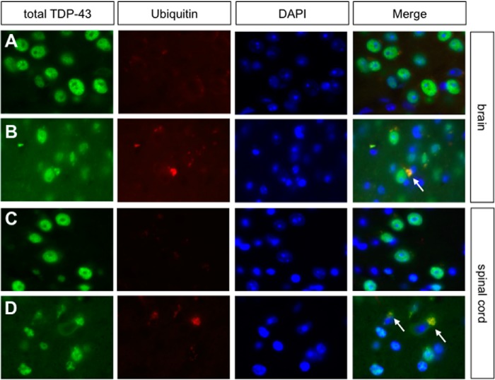
FIGURE 4.
Ubiquitin pathology in brains and spinal cords of 1-year-old mutant mice. A–D, co-labeling of ubiquitin aggregates with total TDP-43 antibody revealing co-aggregation of ubiquitin and TDP-43 in the cytoplasm of brain (B) and spinal cord (D) (white arrows). Co-aggregation was not seen in brain (A) and spinal cord (C) sections of control mice.

