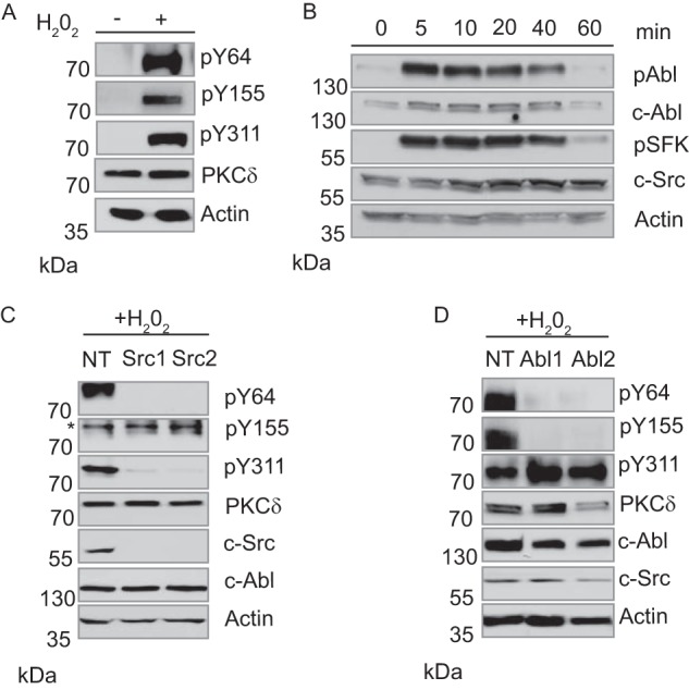
FIGURE 1.

c-Src and c-Abl phosphorylate PKCδ in response to H2O2. A, ParC5 cells were left untreated or treated with 5 mm H2O2 for 10 min. Whole cell lysates were separated by SDS-PAGE and probed using phospho-specific antibodies against PKCδ pY64, pY155, and pY311. To determine loading, membranes were stripped and probed for total PKCδ and actin. B, ParC5 cells were treated with 5 mm H2O2 for the indicated times. Whole cell lysates were resolved by SDS-PAGE and probed using a phospho-specific antibody against the c-Abl activation site (pY412) or the conserved SFK activation site (pY416). Membranes were stripped and re-probed for total c-Abl and c-Src. C and D, ParC5 cells stably expressing either an nontargeting shRNA or two unique shRNAs against c-Src (C) or c-Abl (D) were treated with 5 mm H2O2 for 10 min. Whole cell lysates were resolved using SDS-PAGE and analyzed for PKCδ pY64, pY155, and pY311. Blots were stripped and probed for total PKCδ and actin. Efficiency and specificity of knockdown were determined by probing for c-Src and c-Abl. In C an asterisk denotes the band representing PKCδ pY155. MW, molecular mass.
