FIGURE 2.
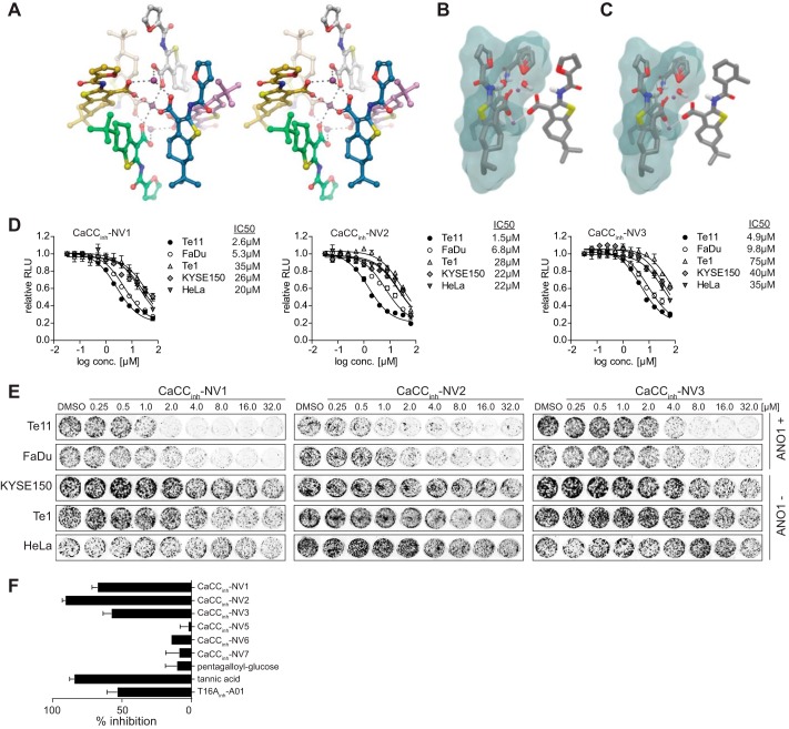
Virtual screening identifies novel inhibitors of ANO1-dependent cell proliferation. A, cross-eyed stereoimage of the crystal structure of CaCCinh-A01. No hydrogen atoms have been added, and water and solvent molecules have been omitted for clarity. Atoms are colored by element, with dark purple, red, blue, and yellow representing calcium, oxygen, nitrogen, and sulfur, respectively. The carbons are colored by molecule for clarity. B and C, crystal structure unit cell of CaCCinh-A01 used to model CaCCinh-A01 analogs. The excluded volume used from the crystal structure unit cell is shown outlined in blue, whereas the modeled compounds (B, CaCCinh-A01; C, CaCCinh-NV2) are shown on the right. The calcium ions are shown in purple. D and E, effect of CaCCinh-NV1–3 on cell viability (D) and colony formation (E) of the indicated cell lines (mean ± S.E. (error bars), n = 4; representative images of stained colonies in a 24-well plate are shown). F, relative inhibition of ANO1 currents in Te11 cells as compared with CaCCinh-A01. The indicated compounds were tested at 30 μm (mean ± S.E., n = 3).

