FIGURE 2.
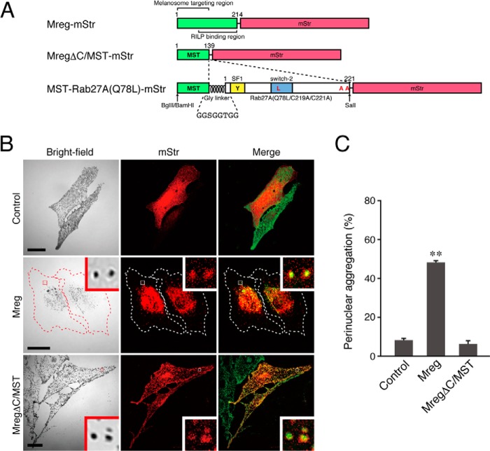
The N-terminal portion of Mreg is necessary and sufficient for melanosome targeting in melanocytes. A, schematic of the Mreg mutants and MST tag used in this study. The N-terminal portion of Mreg is required for its melanosome localization (19, 40), whereas its C-terminal portion binds Rab-interacting lysosomal protein, which forms a retrograde melanosome transport complex (19). MST-Rab27A(Q78L)-mStr was produced by insertion of Gly-linker+Rab27A(Q78L/C219A/C221A) into the site between MST and mStr. BamHI, BglII, and SalI are restriction enzyme sites that were used for the construction of MST-Rab27A(Q78L)-mStr (see “Experimental Procedures” for details). RILP, Rab-interacting lysosomal protein. B, typical images of melanocytes expressing mStr-tagged Mreg mutants and mStr alone (mStr fluorescence images and their corresponding bright-field images). Cells exhibiting perinuclear melanosome aggregation are outlined with a dashed line. The melanosomes in the merged images (right column) are pseudocolored green. The insets show magnified views of the boxed areas. Note that the full-length Mreg with mStr-tag was clearly targeted to mature melanosomes and that Mreg-mStr-expressing cells often exhibited perinuclear melanosome aggregation (center row). MregΔC/MST-mStr, on the other hand, was able to target mature melanosomes without altering peripheral melanosome distribution (bottom row). Scale bars = 20 μm. C, the number of melanocytes exhibiting perinuclear melanosome aggregation expressed as a percentage of the number of transfected melanocytes shown in B. **, p < 0.01; unpaired Student's t test.

