FIGURE 3.
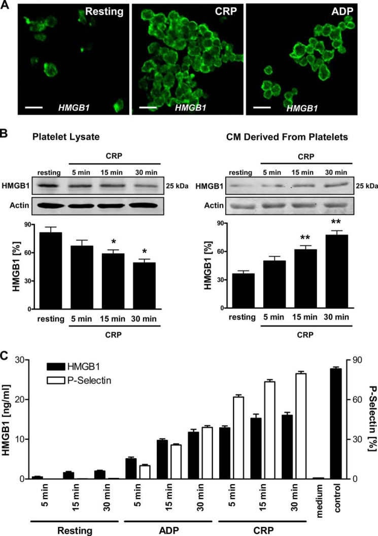
Expression of HMGB1 in platelets and release upon platelet activation. A, expression of intracellular HMGB1 in resting (untreated) and activated (5 μg/ml CRP, 15 min; 50 μm ADP, 15 min) platelets was determined by immunofluorescence staining and confocal laser-scanning microscopy. An HMGB1-specific polyclonal antibody and an Alexa Fluor 488-tagged antibody (green) were used. Representative images of three independent experiments. Scale bar, 5 μm. B, HMGB1 protein levels in lysates and conditioned media derived from resting and CRP-activated platelets (5 μg/ml CRP; 5, 15, and 30 min) were deciphered by Western blot analysis, detecting HMGB1 at 25 kDa. An actin polyclonal antibody served as loading control. Statistical significance (*, p < 0.04; **, p < 0.01) of densitometric analysis of HMGB1 bands (normalized to actin) is indicated. C, HMGB1 levels in conditioned media derived from resting (untreated, 5, 15, 30 min) and activated (50 μm ADP, 5, 15, 30 min; 5 μg/ml CRP, 5, 15, 30 min) platelets were also measured by ELISA. In addition, surface expression of P-selectin (CD62P) on platelets was investigated by staining with a CD62P-specific monoclonal antibody and flow cytometry. Error bars, S.E.

