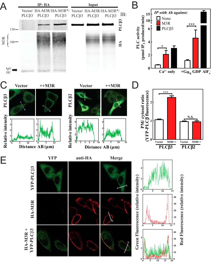
FIGURE 1.
M3 muscarinic receptor stably interacts with PLCβ3. A, HEK293 cells were transfected with PLCβ3 and empty vector or PLCβ3 and 3xHA-M3R with and without stimulation with 100 μm carbachol for 5 min (* indicates treatment with carbachol). Cells were lysed, immunoprecipitated (IP), and immunoblotted (IB) for either PLCβ3 or HA as described under “Experimental Procedures.” Representative Western blots shown were from three or more independent experiments. B, M3R or PLCβ3 was immunoprecipitated (IP) from rat lung lysates and assayed for associated PLC activity as described under “Experimental Procedures.” The data were compiled from four independent assays, each with internal triplicates. C, YFP-PLCβ3 or YFP-PLCβ2 was expressed in CHO cells with empty vector or 3xHA-M3R, and cells were analyzed by live cell confocal microscopy as described under “Experimental Procedures.” Line scans from a to b are shown below each image. D, multiple images treated as in C were analyzed as described under “Experimental Procedures,” compiled, and plotted. E, YFP-PLCβ3, 3xHA-M3R, or YFP-PLCβ3+M3R was expressed in CHO cells. Surface M3R was immunostained in red as described under “Experimental Procedures.” Line scans are shown to the right of each image set. Error bars indicate S.E. *, p < 0.05; ***, p < 0.001; N.S., not significant. Ab, antibody.

