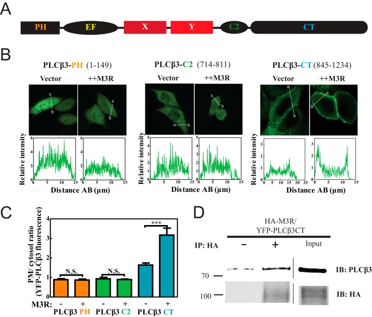
FIGURE 2.
Plasma membrane localization of PLCβ3 C terminus depends on M3R expression and binding. A, primary structure of PLCβ with PH domain (residues 1–147) followed by four EF hands, X and Y catalytic cores, C2 domain (residues 712–809), and CT (residues 845–1234). B, YFP-PLCβ3 fragment constructs were expressed in CHO cells. Either empty vector or 3xHA-M3R was cotransfected. Live cells were analyzed by confocal microscopy as described under “Experimental Procedures.” Line scans from a to b are shown below each image. C, multiple images treated as in B were analyzed as described under “Experimental Procedures,” compiled, and plotted. D, HEK293 cells were cotransfected with YFP-PLCβ3 CT (residues 845–1234) and 3xHA-M3R. Cells were lysed and immunoprecipitated (IP) with or without anti-HA specific antibody. Input lysate (rightmost lanes) and immunoprecipitated (left) samples were immunoblotted (IB) for either PLCβ3 or HA as described under “Experimental Procedures.” Representative Western blots shown were from two independent experiments. Error bars represent S.E. ***, p < 0.001; N.S., not significant.

