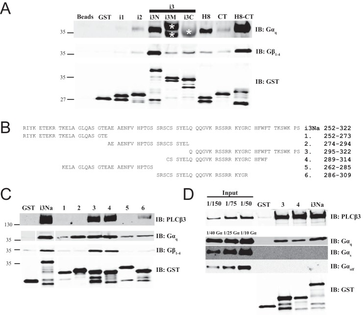
FIGURE 6.
Intracellular loops of M3 muscarinic receptor bind Gαq and Gβγ. A, similar to Fig. 4A, M3R constructs fused to GST were tested for binding to purified Gαq or Gβ1γ2 in a glutathione bead pulldown assay. Results were analyzed by Western blot. Nonspecific recognition for GST fusion proteins by Gαq antibody W082 is indicated by * as shown. Representative Western blots shown were each from three independent experiments. B, within M3Ri3Na (252–322): 1, residues 252–273; 2, residues 274–294; 3, residues 295–322; 4, residues 289–314; 5, residues 262–285; 6, residues 286–309. C, binding site mapping for PLCβ3, Gαq, and Gβγ at M3Ri3N residues 252–322. To map binding sites for PLCβ3 (top), Gαq (middle), or Gβγ (bottom), GST fusion proteins as described in B were used in a pulldown assay. Representative Western blots shown were each from two independent experiments. D, binding of PLCβ3 and different isoforms of G protein α subunits (Gαq, Gαs-long, and Gαolf) to M3Ri3Na fragments 3 and 4 was tested. Results were analyzed by Western blot. Representative Western blots are shown from three independent experiments. Fractions of original input were loaded in the leftmost lanes. Samples were also immunoblotted (IB) with anti-GST antibodies to validate loading equal amounts of protein.

