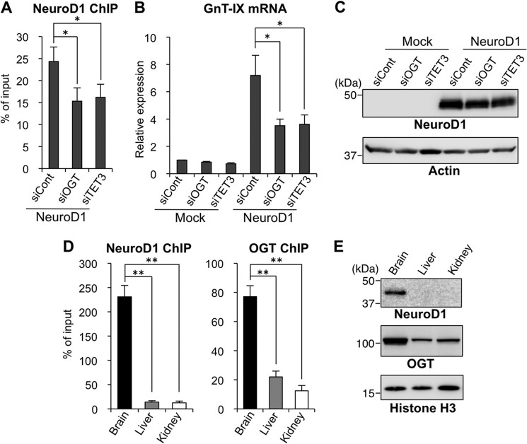
FIGURE 5.
Transactivation of GnT-IX by NeuroD1 is promoted by the OGT-TET3 complex. A, Neuro2A cells were treated with control siRNA (siCont), OGT siRNA, or TET3 siRNA for 24 h and then replated on new dishes. After 24 h, the cells were transfected with the NeuroD1 expression vector followed by a further 24-h culture. Then, ChIP assays were performed with an anti-NeuroD1 antibody. A genomic region around the transcriptional start site of GnT-IX gene, including the NeuroD1 binding site, was analyzed by real-time PCR. The amounts of DNA relative to those of 2% input samples are shown (n = 3, *, p < 0.05, Tukey-Kramer's test). B and C, Neuro2A cells were transfected with siRNA and expression plasmid as in the case of A. Total RNAs were extracted, and the mRNA levels of GnT-IX were quantified and normalized to those of rRNA. The mRNA/rRNA levels relative to those of control siRNA-treated mock sample are shown (n = 3, *, p < 0.05, Tukey-Kramer's test) (B). Cellular proteins extracted after 24 h of NeuroD1-transfection were Western blotted with an anti-NeuroD1 or anti-actin antibody (C). D, chromatin was prepared from brain, liver, or kidney of adult mice, and ChIP assays were performed with anti-NeuroD1 or anti-OGT antibody. A genomic region around the transcriptional start site of GnT-IX gene, including the NeuroD1 binding site, was analyzed by real-time PCR. The amounts of DNA relative to those of 2% input samples are shown (n = 3, **, p < 0.01, Tukey-Kramer's test). E, homogenates of brain, liver, or kidney from adult mice were Western blotted with an anti-NeuroD1, anti-OGT, or anti-histone H3 antibody. All graphs show means ± S.E.

