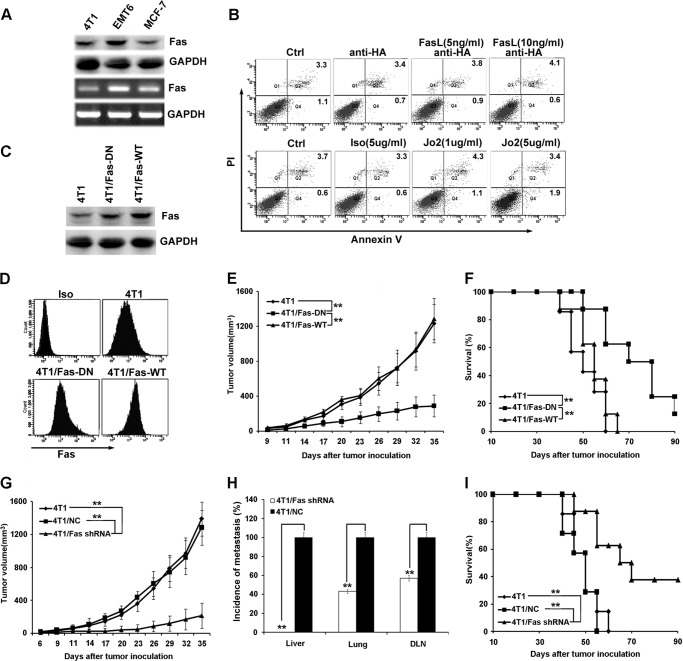
FIGURE 1.
Blockade of Fas signaling in breast cancer cells suppresses tumor growth and metastasis in vivo. A, Fas expression on mouse and human breast cancer cells was detected by Western blot (upper) and RT-PCR (lower). B, susceptibility of 4T1 cells to Fas-induced apoptosis was measured by staining with annexin V-FITC and propidium iodide (PI) after treatment with or without agonistic anti-Fas antibody, Jo2, or cross-linking Fas ligand at the indicated concentrations for 24 h. C and D, expression of Fas in stably transfected 4T1 cell clones (4T1/Fas-WT and 4T1/Fas-DN) was identified by Western blot assay (C) and flow cytometry (D). Iso, isotype. E and F, 5×10⁵ 4T1/Fas-WT, 4T1/Fas-DN, and parental 4T1 cells were inoculated s.c. into the flank of BALB/c mice, then tumor growth and the survival of tumor-bearing mice were monitored and analyzed as described under “Materials and Methods.” G, H, and I, 5 × 10⁵ Fas-silenced 4T1 cell clones (4T1/Fas shRNA), negative control 4T1 cell clone (4T1/NC), and parental 4T1 cells were inoculated s.c. into the flank of BALB/c mice. Tumor growth (G), quantification of the proportion of animals with liver, lung, and DLNs micrometastasis (H), and survival (I) were monitored and analyzed. Data points represent the mean ± S.E. from eight mice per group. Tumor sizes were compared at various time points, and p values are denoted. Survival data were analyzed by log-rank statistics, and the p value is denoted. **, p < 0.01.

