FIGURE 3.
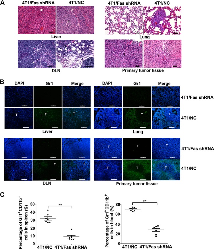
Silencing of Fas expression in breast cancer reduces the mobilization and recruitment of MDSCs in vivo. A–C, 5×10⁵ 4T1/Fas shRNA and 4T1/NC cells were inoculated s.c. into the flank of BALB/c mice; 40 days later, peripheral blood, spleen, liver, lung, DLN, and tumor tissues from tumor-bearing mice were collected. Metastatic tumors in liver, lung, and DLN were detected by HE staining (A). Infiltration and the location of Gr1+ MDSCs in primary tumor tissue or in metastatic tumor tissue in liver, lung, and DLN were measured by immunofluorescence staining (B). The percentage of MDSCs in spleen and peripheral blood (C) was analyzed by FACS as described above. **, p < 0.01. The results represent three independent experiments with similar results; the bar represents 50 μm.

