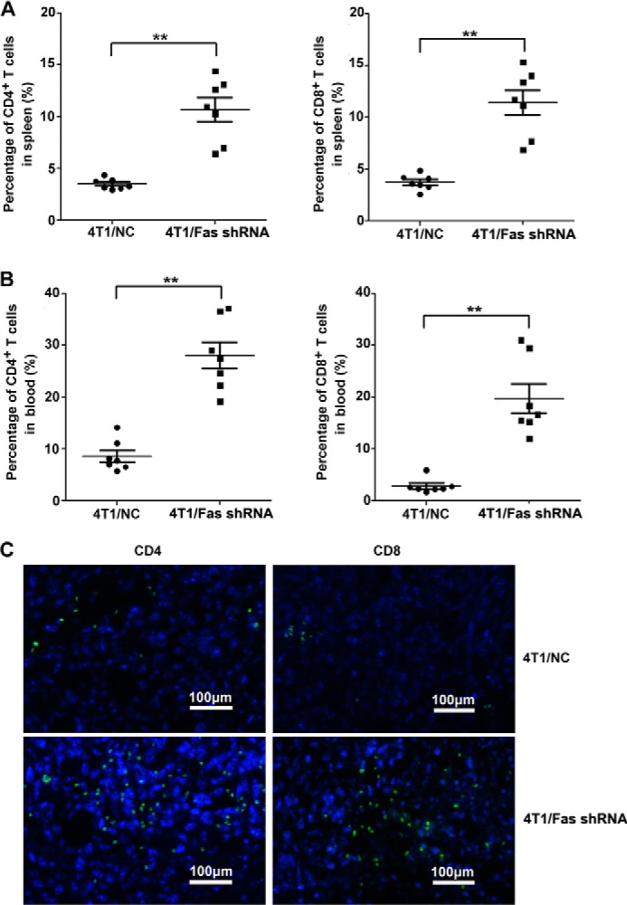
FIGURE 5.

Silencing of Fas expression in breast cancer cells enhances the number of activated T cells in vivo. A–C, 5 × 10⁵ 4T1/Fas shRNA and 4T1/NC cells were inoculated s.c. into the flank of BALB/c mice. 14 days later, peripheral blood and spleen from tumor-bearing mice were collected and stained with FITC-CD3, PE-Cy5-CD4, and APC-CXCR3 for CD3+CD4+CXCR3+ T cells and FITC-CD3 and Percp-CD8 for CD3+CD8+ T cells. The percentage of CD3+CD4+CXCR3+ T cells and CD3+CD8+ T cells in peripheral blood (A) and spleen (B) was analyzed by FACS. Results represent mean value ± S.E. of 3 independent experiments with similar results. *, p < 0.05 and **, p < 0.01. C, infiltrated of CD4+/CD8+ T cells were stained with rat anti-mouse CD8α or rat anti-mouse CD4 antibody followed by staining with Alexa Fluor 488 rabbit anti-rat IgG in tumor tissue as detected by immunofluorescence assay. The bar represents 100 μm.
