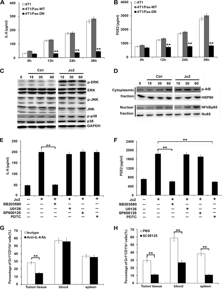
FIGURE 6.
Blockade of Fas signaling in breast cancer cells inhibits proinflammatory cytokine production and MDSC accumulation. A and B, 1 × 10⁶ 4T1/Fas-WT, 4T1/Fas-DN, and parental 4T1 cells were stimulated by 1 μg/ml Jo2 for the indicated times. IL-6 (A) and PGE2 (B) secretion in culture supernatants were detected by ELISA. Data are represented as the mean ± S.D. of three independent experiments. C and D, Western blot analysis of MAPK (C) and NF-κB (D) signal pathways in 4T1 cells cultured in medium alone (Ctrl) or stimulated with Jo-2 for the indicated time. E and F, 1 × 10⁶ 4T1 cells were pretreated with NF-κB inhibitor, pyrrolidine dithiocarbamate (PDTC; 15 μm), JNK/SAPK inhibitor SP600125 (40 μm), p38 MAPK inhibitor SB203580 (30 μm), and MER1/2 inhibitor U0126 (30 μm) for 30 min before Jo2 simulation. IL-6 (E) and PGE2 (F) secretion was detected 24 h later by ELISA. Results are showed as the mean value ± S.D. of the data obtained from three independent experiments. **, p < 0.01. G and H, 5 × 10⁵ 4T1 cells were inoculated s.c. into the flank of BALB/c mice, 125 μg of anti-IL-6 Ab and control isotype (mouse IgG) (G), or COX2 inhibitor SC58125 (5 mg/kg) and PBS (H) were injected intraperitoneally once a day for 14 days. Mice were then sacrificed, and cells were isolated from peripheral blood, spleen, and tumor tissue and stained with FITC-CD11b and PE-Gr1 for analysis of MDSCs by FACS. Experiments were performed three times, and each group contained eight mice. **, p < 0.01.

