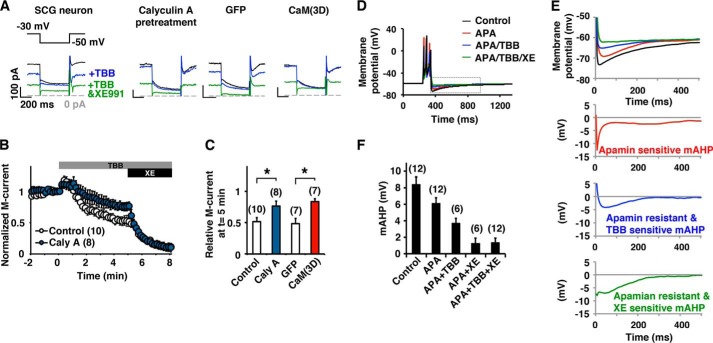
FIGURE 7.
CK2 mediates modulation of the M-current and mAHP in SCG neurons. A, current traces of perforated patch clamp recordings from rat SCG neurons showing that the M-current was partially inhibited by 10 μm TBB and completely inhibited by a combined application of TBB and 10 μm XE991. B, pooled results showing M-current inhibition by TBB and XE991. Responses from untreated control (open symbols) or pretreatment with 10 nm calyculin A (blue symbols) are shown. The gray bar and the black bar indicate the presence of TBB and XE991, respectively. C, summary histogram of current at t = 5 in C showing that calyculin A attenuated TBB-induced suppression. Overexpression of CaM(3D) induced similar attenuated response to TBB (red). *, p < 0.05 by Mann-Whitney test. D, current clamp recordings showing the mAHP in SCG neurons before (black) and after (red) application of 100 nm apamin, subsequent addition of 10 μm TBB (blue), and 10 μm XE991 (green). The resting membrane potential of the cell was kept at −60 mV. mAHP was induced by 100-ms current injection. E, magnified views of the dotted gray box shown in D (top) and dissected components of mAHP in the traces shown. F, summary of incremental decrease of peak mAHP under indicated pharmacological treatments showing that TBB suppressed XE991-sensitive mAHP. Error bars show S.E.

