
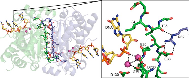
FIGURE 1.
The hydrogen-bonding network at the TREX1 dimer interface. The TREX1 dimer (Protein Data Bank code 2OA8, green/blue) is shown with the extensive side chain-side chain, side chain backbone, and water-bridged (red spheres) hydrogen-bonding network at the interface highlighted. The non-activating Ca2+ ions (pink spheres) and 4-mer ssDNA (yellow sticks) are shown in the active sites. In the box, the Arg-62 residue of one protomer (blue) is shown stretched across the dimer interface, positioning the guanidinium group adjacent to the active site in the opposing protomer (green). The Arg-62 is positioned for interactions with Glu-33 and Thr-85 to correctly orient Ile-84 adjacent to the terminal 3′-nucleotide of the DNA substrate. The metal-coordinating residues (Asp-18, Glu-20, Asp-130, and Asp-200) are indicated. The figure was prepared using PyMOL (Delano Scientific).