
Figure 3.
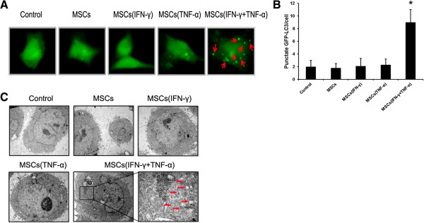
Conditioned medium from the culture of inflammatory cytokine-stimulated MSCs induced autophagy in HCC cells. (A) SMMC-7721 cells were transfected with GFP-tagged LC3; after 24 hours transfection, cells were incubated with contidioned medium collected from MSCs which were pretreated with inflammatory cytokines or not. Images were taken under a fluorescence microscope. Arrows show the punctate GFP-LC3 in the cytoplasm. (B) The number of punctate GFP-LC3 in each cell of SMMC-7721 was counted and at least 100 cells were included for each group. The results were shown as means (±SD) (*p < 0.05). (C) Electron micrographs showing the ultrastructure of SMMC-7721 cells with or without conditioned medium collected from MSCs pretreated with inflammatory cytokines or not. Arrows indicate the autophagic vacuoles in the cytoplasm. Magnification, ×10,000.