
Figure 6.
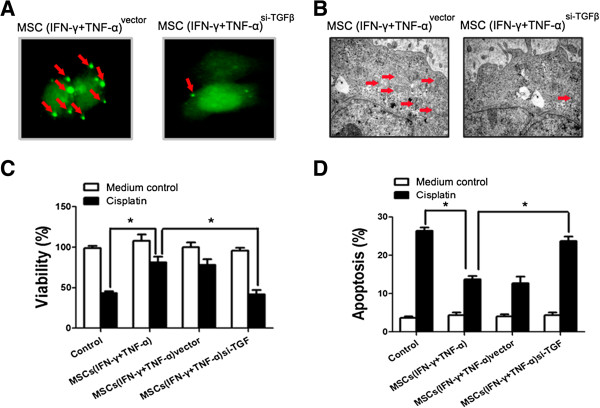
Inhibition of TGF-β expression by MSCs diminished the ability of MSCs in inducing autophagy and chemoresistance in HCC cells. (A) MSCs were transfected with si-TGFβ and then been stimulated by both IFN-γ and TNF-α for 12 hours. The conditioned medium was collected for further investigation. SMMC-7721 cells were transfected with GFP-tagged LC3; after 24 hours transfection, cells were incubated with contidioned medium collected from MSCssi-TGFβ. Images were taken under a fluorescence microscope. Arrows show the punctate GFP-LC3 in the cytoplasm. (B) Electron micrographs showing the ultrastructure of SMMC-7721 cells with or without conditioned medium collected from MSCs pretreated with inflammatory cytokines or not. Arrows indicate the autophagic vacuoles in the cytoplasm. Magnification, ×10000. (C) SMMC-7721 cells (1 × 10⁴/well) were cultured in a 96-well plate with an existence of cisplatin (20 μM) and the conditioned medium collected from MSCssi-TGFβ was added in SMMC-7721 culture medium for 24 hours. MTT was employed to examine the proliferation of SMMC-7721 cells. (D) SMMC-7721 cells were cultured in a 6-well plate with an existence of cisplatin (10 μg/mL) and the conditioned medium collected from MSCssi-TGFβ was added in SMMC-7721 culture medium for 24 hours. PI/Annexin V-FITC assay was used to measure apoptotic SMMC-7721 cells by flow cytometry. (*P < 0.05).