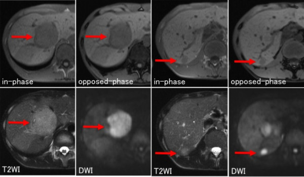
Figure 2.

Abdominal magnetic resonance (MR) imaging. Abdominal magnetic resonance (MR) imaging revealed that the hepatic tumors (segment 5: arrow, segment 6: arrowhead) were slightly hypointense on T1-weighted imaging (WI), slightly hyperintense on T2WI, and hyperintense without an apparent fat component on diffusion-weighted imaging.
