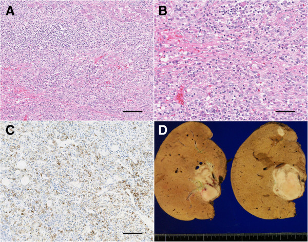
Figure 4.

Histological features of hepatic angiomyolipomas. (A, B) Histological examination revealed the background infiltration of numerous inflammatory cells, including spindle-shaped cells (A: original magnification = 20×, scale bar = 50 μm, B: 40×, scale bar = 100 μm; hematoxylin–eosin staining). (C) The immunohistochemical analysis revealed positive staining for human melanoma black-45 (HMB-45; original magnification = 20×, scale bar = 50 μm). (D) In a gross examination, the cut AML surfaces revealed well-delineated borders and slightly variegated appearances.
