
FIGURE 5.
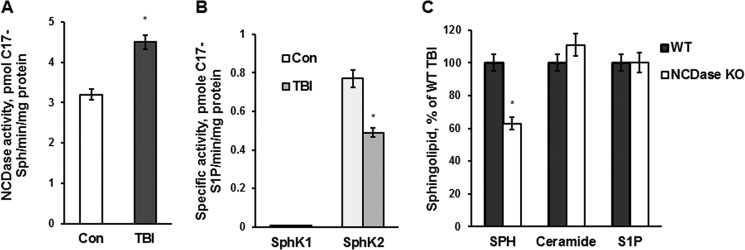
TBI-induced mitochondrial sphingosine accumulation is due to an activation of NCDase and could be partially rescued by knocking down NCDase. Mitochondria were purified from the injured hemisphere of a mouse brain (TBI) and sham-injured mouse brain (Con) at 7 days post-TBI. A, specific NCDase activity was measured with C17-C18-ceramide as a substrate (42), yielding C17-sphingosine (C17-Sph) as described under “Experimental Procedures.” Data are means ± S.E. (error bars); *, p < 0.05; n = 8. B, specific SphK2 and SphK1 activities were measured with C17-sphingosine as a substrate, yielding C17-S1P, as described under “Experimental Procedures.” The specific activities of SphK1 and SphK2 in brain homogenate were 1.10 ± 0.12 and 0.68 ± 0.14 pmol of C17-S1P/min/mg of protein, respectively. Data are means ± S.E.; *, p < 0.05; n = 8. C, cerebral mitochondria were purified from WT and NCDase KO mice at day 7 post-TBI. Sphingolipid content was measured by tandem MS. Data are means ± S.E.; *, p < 0.05; n = 12. Each sample was normalized to its respective total protein levels.