
FIGURE 5.
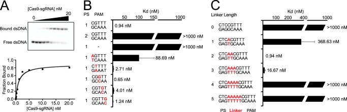
DNA target binding by Cas9. A, a representative gel shift assay for Cas9-sgRNA and the binding curve measured from the assay. B and C, bar graph plotting Kd values for DNA targets with PAM mutations (labeled red) (B) or DNA targets with different linker lengths (labeled red) (C). Average values from at least three replicates are shown, with error bars representing 1 S.D. Targets where binding was not observed are shown with Kd values at the lower limit (> 1000 nm). PS denotes the protospacer sequence.