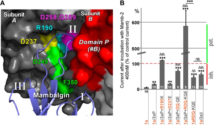
FIGURE 6.
Residues of the β-ball at the bottom of the acidic pocket are necessary for inhibition by mambalgin-2. A, magnification of the modeled interaction between finger II of mambalgin-2 and the acidic pocket of ASIC1a at the interface between subunits A and B. Amino acids of the β-ball putatively involved in the interaction and mutated in subunit B are shown in different colors. Domain P from subunit B is shown in red, and residues Asp-349 and Phe-350 identified in Fig. 3 are also shown. B, bar graph representing the effect of mambalgin-2 (Mamb-2) on different chimeras bearing the domain P of ASIC2a and/or point mutations of key residues from the β-ball of ASIC1a (mapped in A). The number of oocytes analyzed is shown within (or below) each histogram. Data are means ± S.E. (error bars). Statistical comparison is with ASIC1a (*) or ASIC1a/2aP (▵) unless specified. Inhibition (Inh.) or potentiation (pot.) of the current by the toxin is indicated by a red or green bar, respectively.

