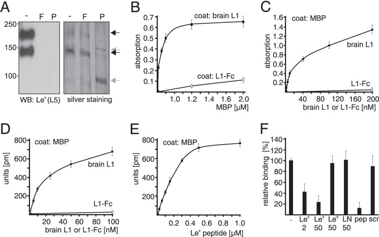
FIGURE 1.
Lex-dependent interaction between L1 and MBP. A, L1 from mouse brain was treated without (-) or with α1–3,4-fucosidase (F) or PNGase F (P) and subjected to Western blot (WB) analysis using Lex-specific antibody L5 or to silver staining to control loading. For ELISA (B and C) and label-free binding assay (D–F), substrate-coated brain L1 (B), L1-Fc (B), or MBP (C–F) were incubated with soluble MBP (B), L1-Fc (C and D), or brain L1 in the absence (C, D, and F) or presence of Lex peptide (E) or a 2- or 50-fold molar excess of Lex, Lea, N-acetyllactosamine (LN), Lex peptide (pep), or scrambled Lex peptide (scr) (F). Mean values ± S.D. from four representative experiments with triplicates are shown for absorption or reflected wavelength shifts relative to values obtained without additions (set to 100%).

