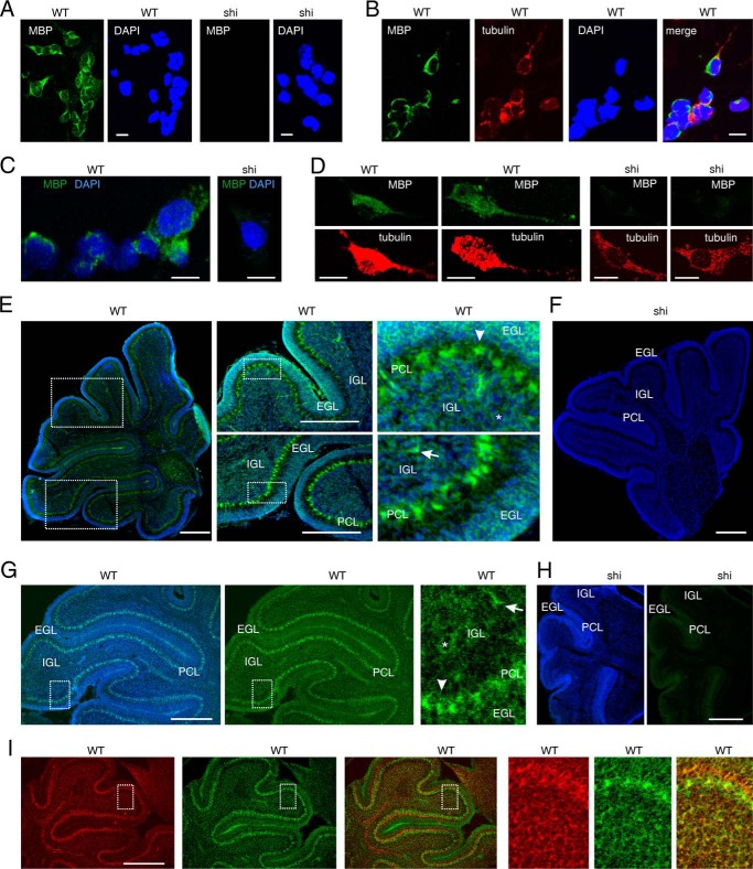
FIGURE 7.
MBP is expressed by cerebellar neurons in vitro and in vivo. Cultured cerebellar neurons (A–D) and cerebellar tissue sections (E–I) from WT and MBP-deficient shiverer (shi) mice were analyzed by in situ hybridization using a probe for the 21.5-kDa MBP isoform (green) (A, B, E, and F) or exon IV-VI-containing MBP isoforms (green) (C, D, and G–I). DAPI (A–D) or bisbenzimide (E–I) was used for nuclear staining (blue) (A–I), and an antibody against the neuronal marker βIII-tubulin (red) was used to identify neurons (B, D, and I). A–I, representative images are shown, and the bars represent 10 μm (A–D) and 300 μm (E–I). EGL, external granular layer; PCL, Purkinje cell layer; IGL, internal granular layer. Granule neurons, interneuron, and Purkinje cells are indicated by asterisks, arrows, and arrowheads, respectively.

