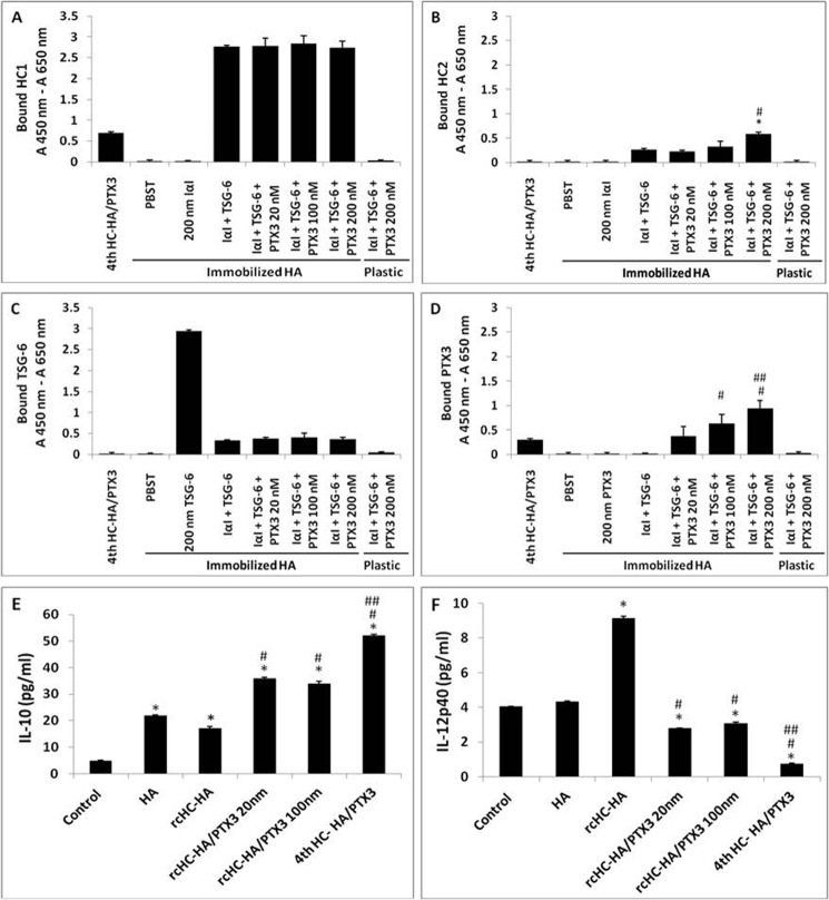
FIGURE 7.
Reconstitution of the HC-HA and HC-HA-PTX3 (rcHC-HA and rcHC-HA-PTX3) complexes in vitro and their effects on macrophage phenotype. A–D, reconstitution of the rcHC-HA and rcHC-HA-PTX3 complexes in vitro. 2 μg of HA, AM fourth HC-HA/PTX3, or PBS control was covalently immobilized on the wells of a Covalink NH 96-well plate. After washing, the wells were blocked with 2% BSA in PBST at room temperature for 1 h. Thereafter, 200 nm IαI, 200 nm TSG-6, or 20–200 nm of PTX3 in PBST containing 5 mm MgSO4 was added into the wells alone or together, as indicated, and incubated for 2 h at 37 °C. The wells were then washed with 6 m GnHCl before ELISA for the bound HC1 (A), HC2 (B), TSG-6 (C), and PTX3 (D) to immobilized HA, respectively. The results represent mean ± S.D. (n = 4). *, p < 0.05 for IαI + TSG-6 + PTX3 (200 nm) versus IαI + TSG-6; #, p < 0.05 for IαI + TSG-6 + PTX3 (100 or 200 nm) versus IαI + TSG-6 + PTX3 (20 nm); ##, p < 0.05 for IαI + TSG-6 + PTX3 (100 or 200 nm) versus IαI + TSG-6 + PTX3 (100 nm). E and F, RAW264.7 cells (3.1 × 10⁴/cm2) were seeded on the aforementioned rcHC-HA and rcHC-HA-PTX3 complexes and treated with 1 μg/ml LPS for 24 h. The protein levels of IL-10 (E) and IL-12p40 (F) were measured in conditioned medium by ELISA. The results represent mean ± S.D. (n = 4). *, p < 0.01 versus control; #, p < 0.01 versus rcHC-HA; ##, p < 0.01 versus rcHC-HA-PTX3 (20 or 100 nm).

