
FIGURE 6.
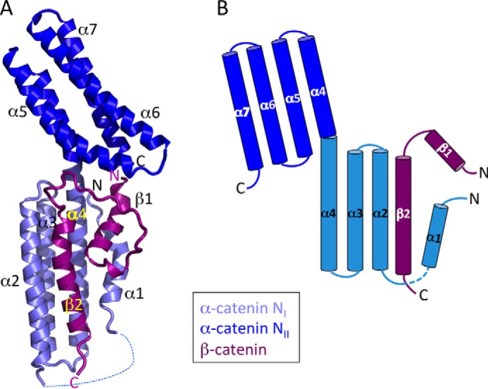
Structure of the complete β-catenin·αN-catenin interface. A, ribbon diagram of the overall structure. Helices β1, β2, α1, and α4 form a small four-helix bundle on the side of the larger NI bundle formed by β2, α2, α3, and α4. The proteins are colored according to the scheme in Fig. 1. B, schematic diagram of the complex showing displacement of the N-terminal αN-catenin helix (α1) from the NI domain by β-catenin.