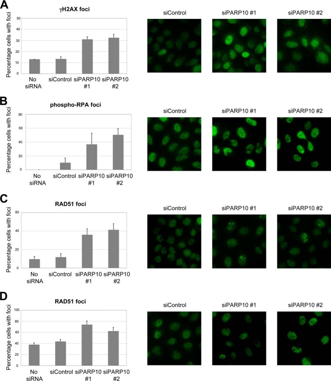
FIGURE 4.
DNA damage accumulates in PARP-deficient cells. UV (100 J/m2)-induced γH2AX foci (A), spontaneous phospho-RPA32 foci (B), spontaneous RAD51 foci (C) as well as CPT (1 μm for 2 h followed by 2 h of recovery) -induced RAD51 (D) foci are shown. HeLa cells were analyzed by immunofluorescence using indicated antibodies. Both non-targeting oligonucleotide (siControl), as well as empty buffer (No siRNA) are shown, as controls. Bars represent the average of at least three experiments in which at least 50 cells were analyzed; error bars are standard errors. Representative micrographs are shown on the left.

